
Heart Ultrasound & Imaging
Echocardiography uses ultrasound technology
to evaluate your heart’s structure, movement,
valves, and pumping function. These noninvasive
tests help our physicians assess cardiovascular
health and identify potential abnormalities.
Echocardiogram (Echo)
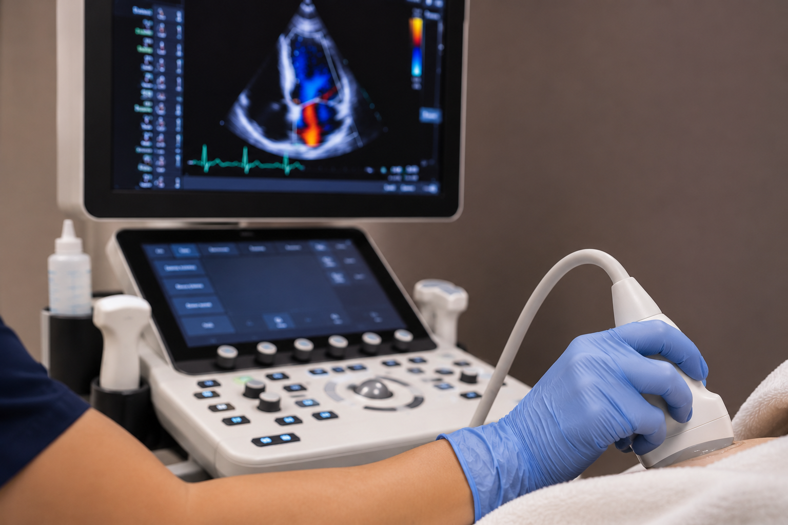
An echocardiogram is a noninvasive ultrasoundexamination that creates moving images of
your heart. It helps evaluate heart chamber size, pumping strength, and heart valve function.
During the examination, a technician moves a small ultrasound device across your chest
while images are recorded. The test typically does not require special preparation.
Stress Echocardiogram
A stress echocardiogram combines ultrasound imaging with exercise to evaluate how your heart functions when working harder.
Images are obtained before and shortly after exercise to assess changes in heart muscle movement and function. Your care team will provide specific preparation instructions before your test.
Cardiac Stress Testing
Cardiac stress testing evaluates how your heart responds to physical activity or medication-induced stress. These tests help physicians assess blood flow to the heart and identify potential signs of coronary artery disease.
Exercise Treadmill Stress Test
An exercise treadmill stress test monitors your heart’s electrical activity while you walk on a
treadmill. The speed and incline gradually increase while your heart rate, blood pressure, and symptoms are monitored.
This test helps evaluate your heart’s response to exercise and may identify abnormalities that are not present at rest.
Myoview Nuclear Stress Test
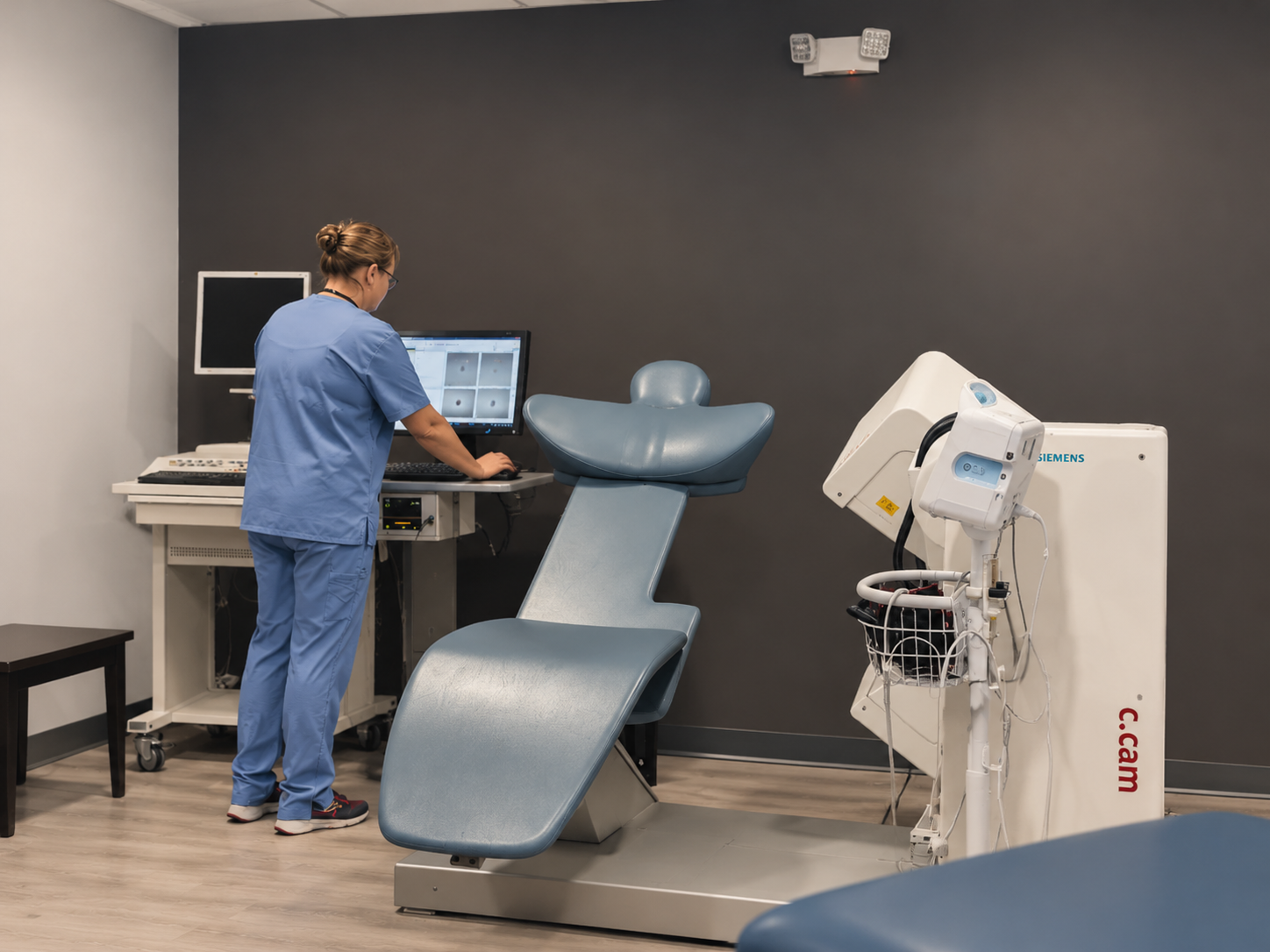
A Myoview nuclear stress test uses a small amount of radioactive tracer and specialized imaging to evaluate blood flow to your heart muscle at rest and during stress.
The stress portion may involve exercise or medication depending on your physician’s recommendation. The resulting images can help identify areas receiving reduced blood flow.
Lexiscan Nuclear Stress Test
A Lexiscan nuclear stress test uses medication rather than treadmill exercise to produce changes in coronary blood flow for imaging.
This test may be recommended when a patient is unable to exercise adequately. Specialized imaging helps evaluate blood flow to the heart muscle.
Heart Rhythm & Monitoring
Heart rhythm testing records the electrical activity of your heart.
These examinations help our physicians evaluate irregular heartbeats, monitor rhythm changes, and identify abnormalities that may not appear during a routine visit.
Electrocardiogram (EKG/ECG)
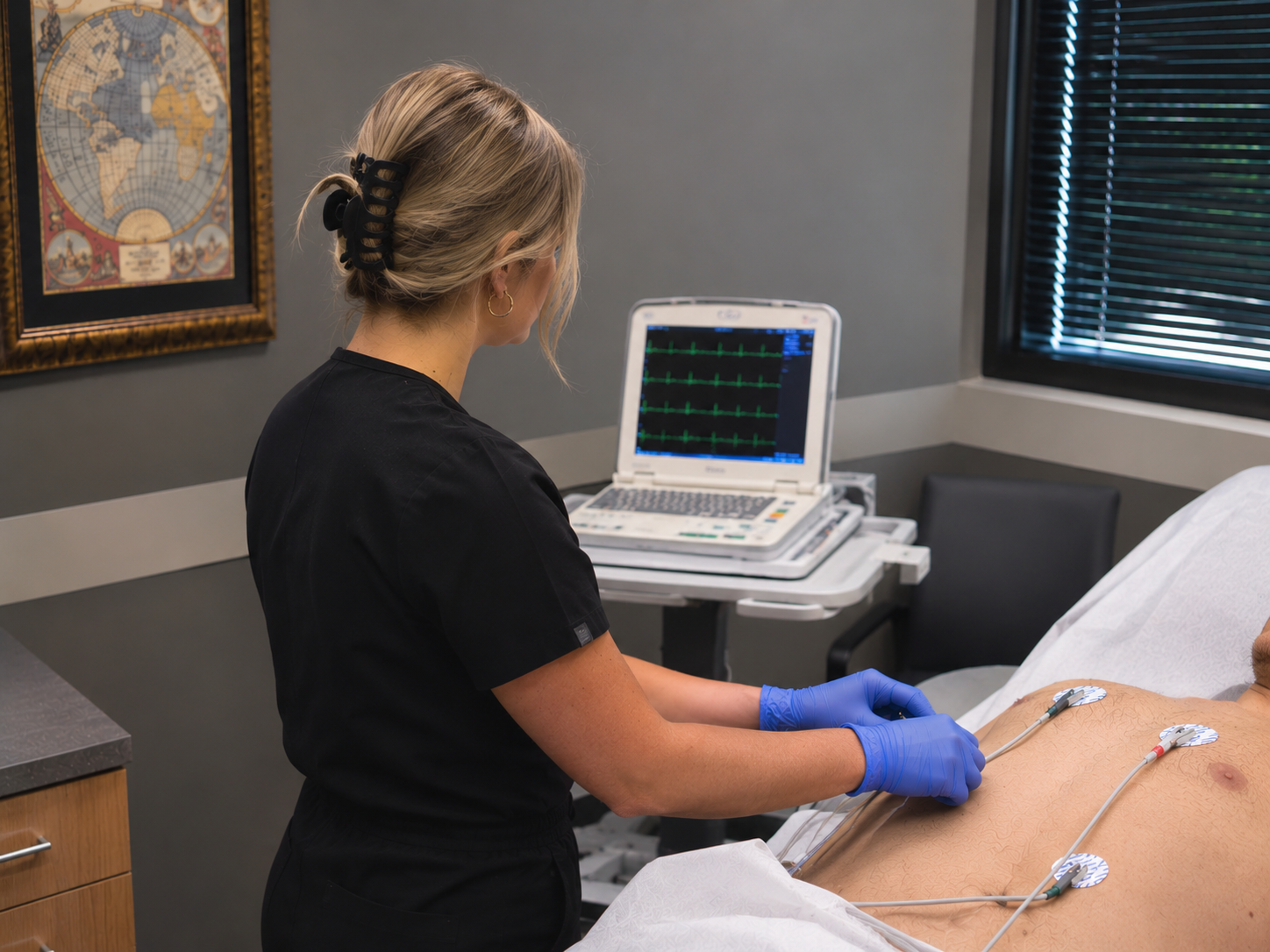
An electrocardiogram is a quick, painless test that records the electrical signals of your heart. Small adhesive electrodes are placed on your chest and limbs.
The test helps evaluate heart rate and rhythm and may detect signs of certain cardiac conditions.
Holter Monitoring
A Holter monitor is a portable device that continuously records your heart’s electrical activity
during normal daily activities.
Extended monitoring can help identify intermittent irregular heart rhythms that may not be
detected during a standard EKG. Your care team will explain how long to wear the device and how to use it.
Vascular Ultrasound Testing
Vascular ultrasound uses sound waves to examine blood flow through arteries and veins. These noninvasive examinations help identify circulation problems, narrowing of blood vessels, blood clots, and venous disease.
Carotid Artery Ultrasound
A carotid ultrasound evaluates blood flow through the carotid arteries in your neck. It can help detect narrowing or plaque buildup that may increase the risk of stroke.
Arterial Ultrasound
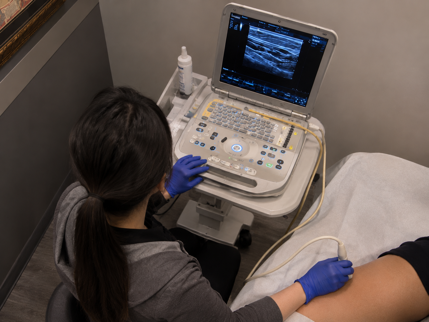
Arterial ultrasound evaluates blood flow through arteries, commonly in the arms or legs. It may help identify narrowing or blockages associated with peripheral artery disease.
Venous Ultrasound & Vein Mapping
Venous ultrasound examines blood flow through the veins and can help identify abnormal vein function or venous reflux.
Vein mapping provides detailed information about vein anatomy and circulation to assist with
treatment planning.
Deep Vein Thrombosis (DVT) Ultrasound
A DVT ultrasound evaluates the deep veins, commonly in the legs, to detect possible
blood clots.
Your physician may recommend this test when symptoms such as unexplained leg swelling, pain, or tenderness occur.
Abdominal & Aortic Ultrasound
Abdominal and aortic ultrasound examines blood vessels within the abdomen, including the abdominal aorta.
This imaging may help identify enlargement, aneurysms, or other abnormalities affecting abdominal blood vessels.
Take the Next Step Toward Better Heart Health
Our cardiovascular team is here to help you understand your testing options and receive personalized, compassionate care.